Nerve Conduction Velocity (NCV) Test
Details
Category: Clinical Engineering
A nerve conduction study (NCS) is a test commonly used to evaluate the function, especially the ability of electrical conduction, of the motor and sensory nerves of the human body. Nerve conduction velocity (NCV) is a common measurement made during this test.
A nerve conduction velocity test measures how quickly electrical impulses move along a nerve. It is often done at the same time as an EMG in order to exclude or detect muscle disorders.
 A healthy nerve conducts signals with greater speed and strength than a damaged nerve. The speed of nerve conduction is influenced by the myelin sheath - the insulating coating that surrounds the nerve. Most neuropathies are caused by damage to the nerve's axon rather than damage to the myelin sheath surrounding the nerve.
The nerve conduction velocity test is used to distinguish between true nerve disorders (such as Charcot-Marie-Tooth disease - also known as hereditary motor and sensory neuropathy (HMSN)) and conditions where muscles are affected by nerve injury (such as carpal tunnel syndrome). This test is used to diagnose nerve damage or dysfunction and confirm a particular diagnosis. It can usually differentiate injury to the nerve fibre (axon) from injury to the myelin sheath surrounding the nerve, which is useful in diagnostic and therapeutic strategies.
During the test, flat electrodes are placed on the skin at intervals over the nerve that is being examined. A low intensity electric current is introduced to stimulate the nerves thus generating nerve impulses.
The Nerve Impulse
The nerve impulse is a wave of cell depolarisation immediately followed by a wave of repolarisation, collectively called an action potential, occurring on the plasma membrane of a nerve fibre. Changes in ion conductances across the nerve fibre membrane are responsible for the initiation and propagation of the action potential. Experimentally, these changes can be the result of electrical current applied through electrodes. Once initiated, an action potential is usually propagated without decrement in amplitude or velocity along the plasma membrane of a nerve fibre.
The velocity or speed of the propagated (conducted) nerve impulse is directly related to the diameter of the nerve fibre and the presence of a myelin sheath. The fastest nerve fibres have large diameters and are myelinated; for example, the motor nerve fibres that supply skeletal muscles. The slowest nerve fibres have small diameters and are unmyelinated; for example, sensory nerve fibre from the stomach.
In the peripheral nervous system, nerve fibres of various diameters and functions (motor and sensory) are bundled together by connective tissue to form nerves. A compound action potential is the sum of all the action potentials occurring in the individual neurons of the whole nerve. The velocity of the compound action potential signal can be a measure and can indicate the state of health of the nerve. Diseases that damage the myelin, destroy neurons, or constrict the whole nerve will decrease the nerve's conduction velocity. However, the nerve conduction velocity may remain normal until late in a disease process as long as a few normal neurons survive. In addition, the nerve conduction velocity reflects conduction of the fastest nerve fibres, usually motor neurons.

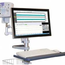
The nerve conduction velocity is determined by recording the motor response of a muscle to the stimulation of its motor nerve at two or more points along the nerve course. The time between stimulation and response is measured and compared to the distance between the point of stimulation and point of response. The velocity at which the resulting electric impulses are transmitted through the nerves is determined when images of the impulses are projected on an oscilloscope or computer screen.
If a response is much slower than normal, damage to the myelin sheath is implied.
If the nerve's response to stimulation by the current is decreased but with a relatively normal speed of conduction, damage to the nerve axon is implied.
The nerve is stimulated, usually with surface electrodes, which are patch-like electrodes (similar to those used for ECG) placed on the skin over the nerve at various locations. One electrode stimulates the nerve with a very mild electrical impulse. The resulting electrical activity is recorded by the other electrodes. The distance between electrodes and the time it takes for electrical impulses to travel between electrodes are used to calculate the nerve conduction velocity. Normal body temperature must be maintained (low body temperature slows nerve conduction).
There is generally minimal discomfort with the test because the electrical stimulus is small and usually is minimally felt by the patient. Often the nerve conduction test is followed by electromyography (EMG) which involves needles being placed into the muscle and you contracting that muscle. This can be uncomfortable during the test, and you may feel muscle soreness at the site of the needles afterwards as well.
NCV is related to the diameter of the nerve and the normal degree of myelination (the presence of a myelin sheath on the axon) of the nerve. Newborn infants have values that are approximately half that of adults, and adult values are normally reached by age 3 - 4.
What abnormal results mean
Most often, abnormal results are caused by some sort of neuropathy (nerve damage or destruction) including:
- Demyelination (destruction of the myelin sheath)
- Conduction block (the impulse is blocked somewhere along the nerve pathway)
- Axonopathy (damage to the nerve axon)
Some of the associated diseases or conditions include:
- Alcoholic neuropathy
- Diabetic neuropathy
- Nerve effects of uremia (from kidney failure)
- Traumatic injury to a nerve
- Guillain-Barre syndrome
- Diphtheria
- Carpal tunnel syndrome
- Brachial plexopathy
- Charcot-Marie-Tooth disease (hereditary)
- Chronic inflammatory polyneuropathy
- Common peroneal nerve dysfunction
- Distal median nerve dysfunction
- Femoral nerve dysfunction
- Friedreich's ataxia
- General paresis
- Lambert-Eaton Syndrome
- Mononeuritis multiplex
- Primary amyloid
- Radial nerve dysfunction
- Sciatic nerve dysfunction
- Secondary systemic amyloid
- Sensorimotor polyneuropathy
- Tibial nerve dysfunction
- Ulnar nerve dysfunction
Any peripheral neuropathy can cause abnormal results, as can damage to the spinal cord and disk herniation (herniated nucleus pulposus) with nerve root compression. There are essentially no risks associated with this procedure.
A NCV test shows the condition of the best surviving nerve fibres and may remain normal if even a few fibres are unaffected by a disease process. A normal NCV test result can occur despite extensive nerve damage. The term NCV often is used to mean the actual test, but this is improper use of the term since velocity is only one measurement out of the entire nerve conduction study. Nerve conduction studies are used mainly for evaluation of paresthesias (numbness, tingling, burning) and/or weakness of the arms and legs. The type of study is determined by the problem.
Some of the common disorders which can be diagnosed by nerve conduction studies include:
- Peripheral neuropathy
- Carpal tunnel syndrome
- Ulnar neuropathy
- Guillain-Barré syndrome
The nerve conduction study consists of the following components:
- Motor NCS
- Sensory NCS
- F-wave study
- H-reflex study
Motor NCS
Motor NCS are performed by electrical stimulation of a peripheral nerve and recording from a muscle supplied by this nerve. The time it takes for the electrical impulse to travel from the stimulation to the recording site is measured. This value is called the latency and is measured in milliseconds (ms). The size of the response - called the amplitude - is also measured. Motor amplitudes are measured in millivolts (mV). By stimulating in two or more different locations along the same nerve, the NCV across different segments can be determined. Calculations are performed using the distance between the different stimulating electrodes and the difference in latencies.
Sensory NCS
Sensory NCS are performed by electrical stimulation of a peripheral nerve and recording from a purely-sensory portion of the nerve, such as on a finger. Like the motor studies, sensory latencies are also measured in ms. Sensory amplitudes are measures in microvolts (uV). The sensory NCV is calculated based upon the latency and the distance between the stimulating and recording electrode
F-wave study
F-wave study uses stimulation of a motor nerve and recording of action potentials from a muscle supplied by the nerve. This is not a reflex, per se, in that the nerve potential travels from the site of the stimulating electrode in the limb to the spinal cord and back to the limb in the same nerve that was stimulated. The F-wave study evaluates conduction velocity of nerves between the limb and spine, whereas the motor and sensory nerve conduction studies evaluate conduction in the limb itself.
H-reflex study
H-reflex study uses stimulation of a nerve and recording the reflexive electrical discharge from a muscle in the limb. This also evaluates conduction between the limb and the spinal cord, but in this case, the afferent impulses (those going towards the spinal cord) are in sensory nerves while the efferent impulses (those coming from the spinal cord) are in motor nerves.
Interpretation of nerve conductions
The interpretation of nerve conduction studies is complex, but in general, different pathological processes result in changes in latencies, motor and/or sensory amplitudes, or slowing of the conduction velocities to differing degrees. For example, slowing of the NCV usually indicates there is damage to the myelin. Another example, slowing across the wrist for the motor and sensory latencies of the median nerve indicates focal compression of the median nerve at the wrist, called carpal tunnel syndrome. On the other hand, slowing of all nerve conductions in more than one limb indicates generalized sick nerves, or generalized peripheral neuropathy. People with diabetes mellitus often develop generalized peripheral neuropathy.
Patient risk
Nerve conduction studies are very helpful to diagnose certain diseases of the nerves of the body. The test is not invasive, but can be a little painful due to the electrical shocks. However, the shocks are associated with such a low amount of electrical current that they are not dangerous to anyone. Patients with a permanent pacemaker or other such implanted stimulators such as deep brain stimulators or Spinal Cord Stimulators must tell the examiner prior to the study. This does not prevent the study, but special precautions are taken.
The nerve conduction study is often combined with electromyography (EMG) where small electrodes on needles are inserted into selected muscles. This is a little painful, but not markedly so. The muscle will be sore at the site of the needle.
Compiled by JD Sandham IEng MIET MIHEEM
Sources:
http://www.webmd.com/hw/brain_nervous_system/hw213852.asp
http://en.wikipedia.org/wiki/Nerve_conduction_study
http://www.nlm.nih.gov/medlineplus/ency/article/003927.htm
http://www.biopac.com/bslprolessons/h03/bslproh03.htm
RECENT ARTICLES
An electroencephalogram (EEG) is a test that looks at the function of the brain. The brain works by a series of nerve impulses, which cause electrical signals. These signals (also called brainwaves)...
Your heart's electrical system controls all the events that occur when your heart pumps blood. The electrical system also is called the cardiac conduction system. The heart test called an ECG...
Electromyography (EMG) is an electro-diagnostic medicine technique for evaluating and recording the electrical activity produced by skeletal muscles. The EMG is performed using an instrument called...

Physiological responses to Action Potential Stimulation The aim of this article is to explain the how Action Potential Stimulation (APS) therapy affects the physiology (especially the...